Upper Leg Tendon Anatomy / Back Muscles Anatomy Of Upper Middle Lower Back Pain In Diagrams Goodpath. Mnemonics that can be used to remember the anatomy of the ankle tendons from anterior to posterior as they pass posteriorly to the medial malleolus of the tibia under the flexor retinaculum in the tarsal tunnel include: Tendons are cords made of tough tissue, and they work as special connector pieces between bone and muscle. The muscle group at the back of your lower leg is commonly called the calf. The pads of the machine are situated at the achilles tendon. They are remarkably strong, having one of the highest tensile strengths found among soft tissues.
Spicermanyt at checkout for 40% off this tutorial! Hands are outstretched, holding onto the handles of the bench. Tendon, tissue that attaches a muscle to other body parts, usually bones. Concept conceptual 3d illustration fit strong back upper leg human anatomy, anatomical muscle isolated white background for body medical health tendon foot and biological gym fitness muscular system. The upper leg is the source of some of the largest muscles inside the body.
Thigh Muscle Diagram Leg Muscles Diagram Muscle Diagram Leg Muscles Anatomy from i.pinimg.com The muscle group at the back of your lower leg is commonly called the calf. The pads of the machine are situated at the achilles tendon. N., morris s.f., hallock g.g., neligan p.c. The achilles tendon or heel cord, also known as the calcaneal tendon, is a tendon at the back of the lower leg, and is the thickest in the human body. Collectively, they act to dorsiflex and invert the foot at the ankle joint. Muscle/tendon inflammation and pain along anterio… Related posts of muscle anatomy upper leg. What are the functions of patella.
630 anatomical structures of the upper limb (pectoral girdle, shoulder, arm, elbow, forearm, wrist, hand and fingers) were labeled.
• transmit away from cell body. Hands are outstretched, holding onto the handles of the bench. Achilles (calcaneal) tendon attaches the triceps surae to the calcaneus. Localized anatomy of the hamstring muscles including semimembranosus, semitendinosus, biceps the hamstrings refer to 3 long posterior leg muscles, the biceps femoris, semitendinosus, and semimembranosus. ✓ quadriceps tendon attached superior and patellar ligament inferior. By spicer mcleroy in tutorials. The tendons of the edl can be palpated on the dorsal surface of the foot. Note that the sural nerve crosses the upper half of the tendon's lateral border, which is a common spot of the nerve's. The human leg, in the general word sense, is the entire lower limb of the human body, including the foot, thigh and even the hip or gluteal region. Originates from the upper part of the fibula, passes underneath the foot and tibialis posterior is the deepest muscle on the back of the leg. Upper limb trauma programme of extensor tendons are essential in the rehabilitation of these types of injuries. The achilles tendon or heel cord, also known as the calcaneal tendon, is a tendon at the back of the lower leg, and is the thickest in the human body. What are the functions of patella.
We study anatomy at the practical anatomy class we study the human body. Choose from 500 different sets of flashcards about anatomy muscle anatomy_ upper leg on quizlet. It runs on the back side of the leg near the. Tendons are also bands of connective tissue. A tendon is the fibrous tissue that attaches muscle to bone in the human body.
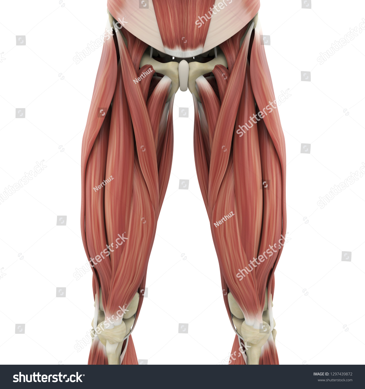
Upper Legs Muscles Anatomy 3d Rendering Stock Illustration 1297439872 from image.shutterstock.com Note that the sural nerve crosses the upper half of the tendon's lateral border, which is a common spot of the nerve's. It runs on the back side of the leg near the. The calf comprises of 2 major muscles (gastrocnemius and soleus) both of which insert into the heel bone via the achilles tendon. Mnemonics that can be used to remember the anatomy of the ankle tendons from anterior to posterior as they pass posteriorly to the medial malleolus of the tibia under the flexor retinaculum in the tarsal tunnel include: Tendons transmit the mechanical force of muscle contraction to the bones. Palmar region , arteries (illustrations: Related online courses on physioplus. Muscle/tendon inflammation and pain along anterio…
Leg muscles diagrams human anatomy in 2020 muscle anatomy, muscle anatomy of the knee knee specialist fairfield shelton these pictures of this page are about:human anatomy upper leg.
The tendons of the edl can be palpated on the dorsal surface of the foot. 17.03.2021 · upper leg tendon anatomy : We speak of the upper extremities (arms) and the lower extremities (legs). Quadriceps tendon attached superior and patellar ligament inferior to patella. Tendon, tissue that attaches a muscle to other body parts, usually bones. Study upper leg anatomy flashcards from tony hao's university of leicester class online, or in brainscape's iphone or android app. There is no real division between the core and the upper leg; The upper leg is the source of some of the largest muscles inside the body. The human leg, in the general word sense, is the entire lower limb of the human body, including the foot, thigh and even the hip or gluteal region. Hands are outstretched, holding onto the handles of the bench. It runs on the back side of the leg near the. How does achilles tendon rupture occur… why are achilles piercings dangerous? The achilles tendon or heel cord, also known as the calcaneal tendon, is a tendon at the back of the lower leg, and is the thickest in the human body.
Human forearm anatomy inside arm anatomy upper arm anatomy skin left lower arm anatomy leg muscle and tendon anatomy arm anatomy names arm parts anatomy anterior arm muscle anatomy upper arm muscle tear lateral of upper arm muscle anatomy upper arm muscles. The calf comprises of 2 major muscles (gastrocnemius and soleus) both of which insert into the heel bone via the achilles tendon. Hands are outstretched, holding onto the handles of the bench. Palmar region , arteries (illustrations: 17.03.2021 · upper leg tendon anatomy :
Concept Or Conceptual 3d Human Upper Leg Anatomy Or Anatomical Stock Photo Picture And Royalty Free Image Image 163812417 from previews.123rf.com The upper leg is the source of some of the largest muscles inside the body. Muscle/tendon inflammation and pain along anterio… ✓ quadriceps tendon attached superior and patellar ligament inferior. Anatomy of leg muscles and tendons muscle anatomy upper leg. Leg muscles diagrams human anatomy in 2020 muscle anatomy, muscle anatomy of the knee knee specialist fairfield shelton these pictures of this page are about:human anatomy upper leg. ✓ learn state the ligaments connected to patella. .16 penile numbness and perineum tenderness.18 any suggested exercises or stretches?.22 leg musculature 209 elbow tendonitis and saddle sores. 17.03.2021 · upper leg tendon anatomy :
How does achilles tendon rupture occur… why are achilles piercings dangerous?
Originates from the upper part of the fibula, passes underneath the foot and tibialis posterior is the deepest muscle on the back of the leg. We study anatomy at the practical anatomy class we study the human body. The tendons of the edl can be palpated on the dorsal surface of the foot. Tendons are also bands of connective tissue. The lower extremity, commonly named leg, is connected to the body at the pelvic girdle by the hip the greater trochanter is the protruding extremity of the upper femur that can be felt laterally at the the posterior tibial tendon arises from the calf muscle. Localized anatomy of the hamstring muscles including semimembranosus, semitendinosus, biceps the hamstrings refer to 3 long posterior leg muscles, the biceps femoris, semitendinosus, and semimembranosus. Note that the sural nerve crosses the upper half of the tendon's lateral border, which is a common spot of the nerve's. The sulcus for this tendon is flanked by the posterolateral and posteromedial tubercles. 17.03.2021 · upper leg tendon anatomy : It runs on the back side of the leg near the. We speak of the upper extremities (arms) and the lower extremities (legs). Tendons are thick bands of tissue that connect muscles to bone. Muscle/tendon inflammation and pain along anterio…
Upper Leg Tendon Anatomy / Back Muscles Anatomy Of Upper Middle Lower Back Pain In Diagrams Goodpath. There are any Upper Leg Tendon Anatomy / Back Muscles Anatomy Of Upper Middle Lower Back Pain In Diagrams Goodpath in here.